


Are Dental X-rays Safe?
Dental X-rays play an essential role in contemporary dentistry by enabling dentists to identify and address oral issues promptly and efficiently. These diagnostic visuals offer crucial information about the well-being of our teeth, bones, and nearby tissues.
Nevertheless, many people continue to harbour apprehensions about dental X-ray safety, especially concerning potential radiation exposure hazards. That’s why it’s essential to understand the safety aspects associated with dental X-rays, as this knowledge empowers us to make educated choices when it comes to our oral health care.
Understanding the Safety of Dental X-rays
Dental X-rays serve as a standard diagnostic instrument used by dentists to evaluate our oral health and identify potential concerns that might not be apparent during a routine checkup. However, while X-rays have indeed transformed dentistry, safety concerns have also emerged among their patients.
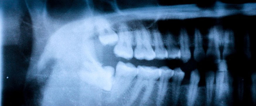
Before undergoing an X-ray, it's crucial to comprehend the nature of dental X-rays and their functioning - X-rays are a type of electromagnetic radiation capable of penetrating various substances, including human tissue. Dental X-rays employ a concentrated radiation beam to obtain images of teeth, bones, and adjacent tissues, and these images assist dentists in detecting issues such as tooth decay, infections, gum disease, and irregularities in our oral structure.
Minimizing Radiation Exposure
The main concern for most individuals when it comes to dental X-rays is the potential risk associated with radiation exposure.
Ionizing radiation, utilized in X-rays, has the potential to harm living tissues and cells. However, it's crucial to recognize that dental X-rays emit very minimal amounts of radiation, particularly when compared to other medical imaging procedures such as CT scans or diagnostic X-rays for other body parts.
Dentists generally take precautions to ensure their patients are exposed to the lowest possible amount of radiation needed to obtain the necessary diagnostic data. Contemporary dental X-ray machines are designed to reduce radiation exposure by employing collimators, lead aprons, and thyroid shields to safeguard areas of the body not being imaged.
Moreover, digital X-ray systems have supplanted traditional film-based techniques, decreasing the exposure time and radiation required to capture an image.
The radiation dosage from dental X-rays is typically measured in millisieverts (mSv), a unit that calculates the amount of radiation absorbed by the body. On average, a standard dental X-ray might deliver a dose of approximately 0.005 mSv, which is significantly lower than the natural background radiation exposure a person receives from the environment annually (roughly 2-3 mSv).
The Benefits of Dental X-rays
Dental X-rays allow dentists to detect oral health issues that may not be visible to the naked eye, facilitating early identification and treatment of oral conditions. This helps prevent the progression of dental problems, decreases the necessity for more invasive procedures, and ultimately leads to improved oral health outcomes.
In some instances, dental X-rays are essential tools for precise diagnosis and treatment planning, particularly for complex procedures like root canals, dental implants, and orthodontic treatments.
The safety of dental X-rays is further supported by guidelines and regulations established by various national dental and radiology organizations. These organizations set standards and recommendations for the proper use of dental X-rays, and all licensed dentists follow these guidelines to ensure patient safety and minimize unnecessary radiation exposure.
Generally, the risk associated with dental X-rays is extremely low for the overall population. However, certain individuals may be more vulnerable to radiation effects - for example, pregnant women are typically advised against routine dental X-rays unless absolutely essential. In such situations, dentists implement additional precautions, such as using lead aprons and thyroid shields, to protect the developing fetus from radiation exposure.
Patients with specific medical conditions or those receiving radiation therapy may also need particular considerations regarding dental X-rays.
Are Dental X-Rays Safe?
Dental X-rays are generally deemed safe since the radiation emitted during the procedure is minimal, particularly when compared to other medical imaging techniques. Dentists implement precautions to reduce your radiation exposure during appointments, and the advantages of dental X-rays for diagnosis and treatment planning surpass their potential risks.
Dental X-rays offer essential diagnostic data that is vital for detecting oral issues that may not be visible to the naked eye. They enable dentists to identify tooth decay, infections, gum disease, and irregularities in your oral structure. Early detection through X-rays can impede the progression of dental problems, diminish the necessity for more invasive procedures, and promote improved oral health outcomes.
Are There Any Risks Associated with Dental X-Rays?
While the risks linked to dental X-rays are minimal, it's important to take individual circumstances into account. Pregnant women, for instance, are generally advised against routine dental X-rays unless absolutely essential, and in such situations, dentists will implement extra precautions to shield the developing fetus from radiation exposure.
Patients with specific medical conditions or those undergoing radiation therapy may also need particular considerations regarding their dental X-rays to ensure their safety.
Dentists employ numerous precautions to reduce your radiation exposure during dental X-rays. Contemporary X-ray machines are designed to concentrate the radiation beam on the area of interest, minimizing scatter radiation, and lead aprons and thyroid shields are also utilized to protect body regions not being imaged. Furthermore, digital X-ray systems have replaced traditional film-based techniques, decreasing exposure time and radiation requirements.
How Often Should Dental X-Rays Be Taken?
The frequency at which you need dental X-rays depends on various personal factors, such as your age, oral health history, and your likelihood of dental issues. Generally, adults with good oral health may require X-rays every 1-2 years, while those with a history of dental problems or undergoing treatment might need them more often.
Dentists use their professional judgment to decide the suitable timing and frequency of dental X-rays for each patient. In some instances, alternative imaging methods like cone-beam computed tomography (CBCT) or ultrasounds may be employed as a substitute for dental X-rays, although these techniques are typically reserved for specific cases where extra information is needed.
Overall, dental X-rays are widely considered safe and can offer valuable diagnostic data for your dentist. The precautions your dental professional takes, combined with modern technological advancements and adherence to guidelines, ensure that you are exposed to the minimum amount of radiation necessary for your health, and the benefits of dental X-rays greatly surpass their potential risks.
It’s important for you as a patient to discuss any concerns or questions you may have with your dentist, who can provide you with personalized information and address your individual needs and circumstances. At Maple Dental Health, our team of professionals is here for you to help you get the highest level of care possible for the results you deserve. Reach out to a member of our team today to schedule your consultation.